
Bones on Demand: Using 3D models in archaeological teaching and learning
Handling ancient remains in the field or lab is integral to archaeology. So why use digital reconstructions? In this blogpost, Rachel Schats will share her experiences with creating and using 3D models and will discuss the potential benefits for archaeological education.
3D visualisations in archaeological teaching: Why bother?
Archaeology is about people and the lives they lived. Central to the field is the detailed study of artefacts (e.g., tools, jewellery, and pottery) and ecofacts (e.g., plants, wood, and human and animal bones). The physical remnants past people leave behind offer key insights into the development and nature of societies. These objects and remains are therefore also at the core of our teaching at the Faculty of Archaeology. Traditionally, archaeological students are exposed to various types of materials and remains during their studies and practical exams in which they correctly have to identify stones and pottery, but also organic remains such as pollen and bones are common. While 3D visualisations and digital tools are often during archaeological excavations and research, this technology has not yet made its way into the formal education at our Faculty. While it is unlikely that digital reconstructions will have the same educational value as handling ancient remains, the use of 3D visualisations in teaching and learning can have many benefits, both in online and in-person teaching, and not just for the archaeological field.
The use of three-dimensional visualizations in educational programmes has already become commonplace in several fields. They have been used extensively in medical education in addition to physical models and examples (Yammine and Violato 2015), but these digital tools have also been used successfully in construction and chemistry education (see for example Perdomo et al. 2005; Abdinejad et al. 2021). In medical teaching, the 3D models allow students to study anatomy outside of the laboratory, but also provide the opportunity to practice medical procedures without actual patients having to be involved (Vernon and Peckham 2009). A survey showed that while students did not support the complete replacement of physical specimens with digital reconstructions, those that supplemented their training with three-dimensional visualisations found them enjoyable to use and they performed significantly better on examinations (Vernon and Peckham 2009).
For archaeological education, the use of 3D visualization could provide similar benefits. As with medical teaching, real specimens are unlikely to be completely replaced, but incorporating virtual models in the traditional laboratory education will allow students to engage, manipulate, and practice with archaeological finds wherever they are. Additionally, with the large student numbers it is difficult for all students to see or study an object at the same time, a digital copy would ensure more and continued access within and outside the classroom. Moreover, certain archaeological objects and materials can be very fragile and/or degrade quickly (e.g., wood), or are so unique that only the lucky few have access. Thus, having access to digital reconstructions will likely increase the study possibilities of the students as well as diversify the objects and materials that can be included in the curriculum. With this in mind, prof. Marie Soressi, Martina Revello Lami, and I started a pilot project (SALTSWAT funded by CfI) to digitize parts of the physical reference collection at the Faculty of Archaeology and to study how this can best be used in education. This endeavour is being continued as part of my LTA project. In this blog, I would like to discuss the pilot project focusing on the process of digitization, the implementation into education, and the initial student feedback to give an insight into the necessary tools and the benefits 3D models can have for students and staff in teaching and learning.
A case study: digitising archaeological human bones
My field of research is human skeletal remains, which, like medical teaching, has a strong focus on anatomy, but also aims to investigate all other aspects of a skeleton, such as sex, age-at-death, and pathological conditions to learn about past populations. While the LTA project will have a much broader focus, for the initial pilot project, I chose to specifically focus on creating models that could help with the estimation of sex, one of the most important aspects of archaeological skeletal analysis. Books and physical models are available to students to study with, yet, they often struggle with understanding the features and the sometimes subtle differences between the skeletons of males and females.
Step 1: Selecting the specimens
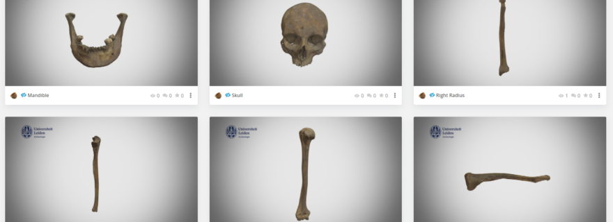
As these are the most sexually dimorphic parts of the skeleton, I selected several complete crania, mandibles, and pelvic bones from archaeological individuals housed in the Faculty of Archaeology that were convincingly male or female. I chose those individuals whom I felt showed all traits clearly and as such could be very useful for students when trying to understand the features, but could also be used during the estimation of sex of an unknown individual, as a digital reference manual.
Step 2: Digitising the bones
The selected bones were digitised by Alicia Walsh, MA who was hired on the project using three different methods: Structure from Motion (also known as photogrammetry), which is a 3D modelling technique that produces spatial 3D point clouds from overlapping 2D images, The NextEngine Ultra HD Scanner, which is a desktop laser scanner, and the Artec Space Spider, based on blue light technology. The photogrammetry approach is relatively easy and all you need is a good camera. The NextEngine and Artec scanners result in more accurate models, but are more expensive, particularly the Artec Space Spider (~€20.000).

Step 3: Viewing the 3D models
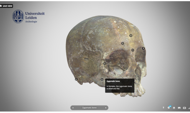
The 3D models can be studied using viewers which are available on most newer devices. Yet, the 3D files are large and therefore not very easy to share. This complicates the direct implementation in education. Moreover, just sharing the files with the students would then assume that all have access to computers able to deal with these types of files. To overcome this, we decided to use the Sketchfab, a commercial company that delivers an online viewer and repository for 3D images and models. While there are downsides to using a commercial platform, the accessibility of the models is excellent. Moreover, Sketchfab allows the models to be annotated which offers the opportunity to give additional explanations about certain features or traits in the models, as can be seen in the image above.
Step 4: Implementation in education
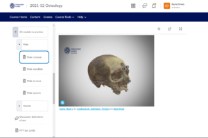
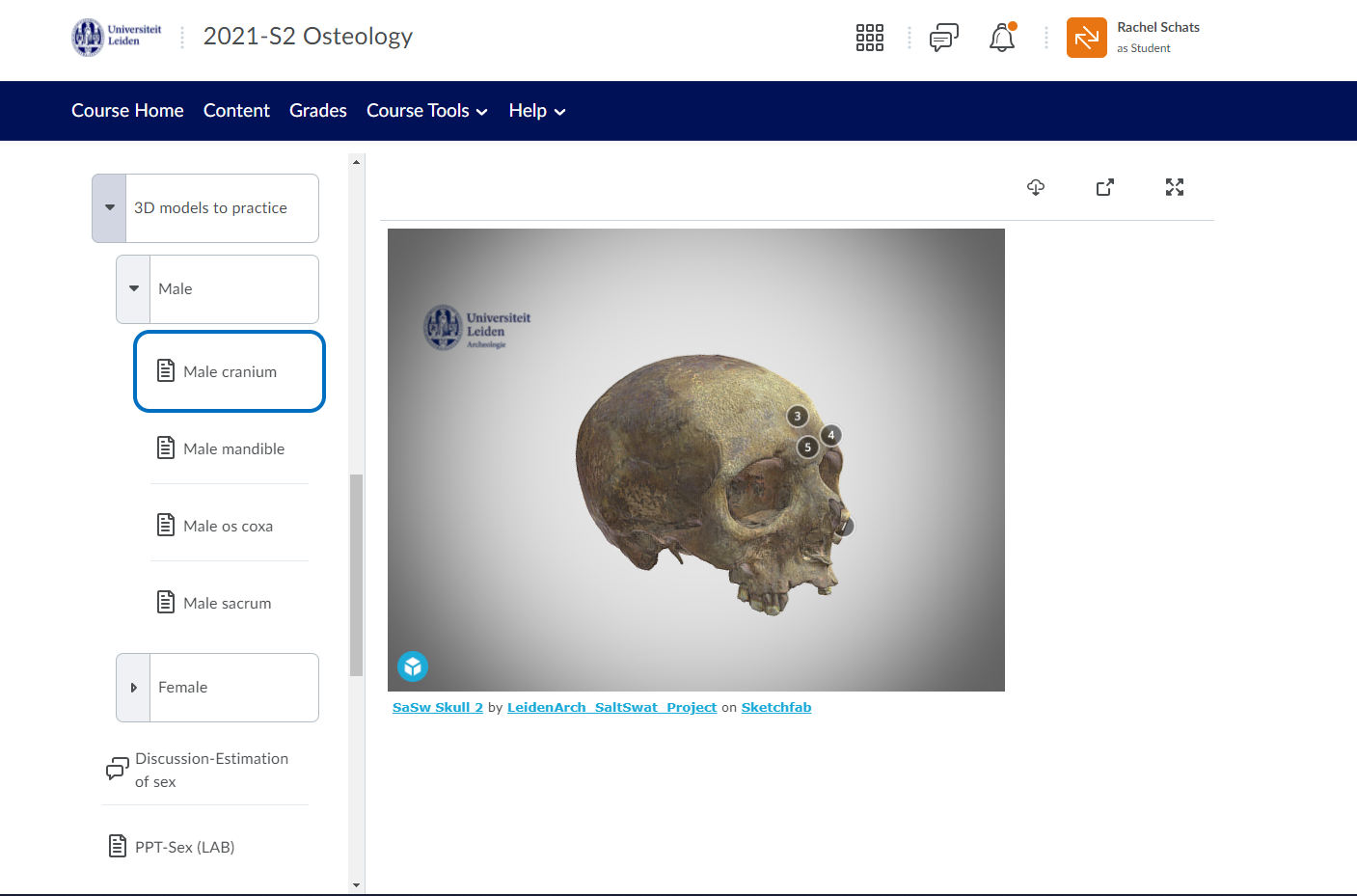
Once the models are uploaded to Sketchfab, they can be directly embedded into the Learning Management System, in this case, Brightspace. This allowed the 3D models to be directly part of the course without the need for the students to access/buy additional software or navigate other websites. The students could access the models at home for independent study, but also in class on their laptops as reference material. During the first lockdown when teaching was taking place online, the 3D models were used as a replacement for physical models and to test the skills of the students.
Step 5: Student feedback
No formal evaluation of their experiences was executed in this pilot project, but during the contact hours, the students expressed they found 3D models a welcome addition to study with at home for the practical test. Especially the siding of bones (i.e. determine if they are left or right), could be practised with these models, which is normally something only possible in the lab. Others also used the models in the lab as reference material. The formal evaluations of the class during lockdown did show that, although the students were happy that they were able to engage with the material in a digital form, 3D models cannot replace the real specimens.
3D models: Worth the effort?
Although more thorough and formal evaluations of the use of 3D models has to be done, preliminary results suggest that students appreciate the addition of digital reconstructions to their education, specifically to study with at home and in class. Especially when practical assessment is part of the course, it is beneficial for the students to have access to the material whenever they want. For me as a teacher, I was very happy to use these models as I was able to select the best ones and give exactly the appropriate amount of information. This is often not the case with physical plastic models we buy commercially or 2D images in books. It is, however, a substantial time (and therefore money) investment to create the digital visualisations. Although photogrammetry can be done relatively cheap (and it makes a great internship for a student), it is a labour-intensive process with a steep learning curve. Yet, the models that we have created are now available for many years to come and can be easily shared with other instructors or interested parties. Moreover, in a world that is very hybrid, we have also been able to use them for outreach activities with the general public as well as during online recruitment sessions. In sum, creating good and informative 3D models is a substantial investment with regard to time and money, but I feel it is absolutely worth the effort.
Further reading
Abdinejad, Maryam, Borzu Talaie, Hossain S. Qorbani, and Shadi Dalili. 2021. Student Perceptions Using Augmented Reality and 3D Visualization Technologies in Chemistry Education. Journal of Science Education and Technology 30(1): 87–96. DOI: 10.1007/S10956-020-09880-2/FIGURES/13.
Perdomo, Jose L., Mohd Fairuz Shiratuddin, Walid Thabet, and Ashwin Ananth. 2005. Interactive 3D visualization as a tool for construction education. ITHET 2005: 6th International Conference on Information Technology Based Higher Education and Training, 2005 2005. DOI: 10.1109/ITHET.2005.1560307.
Vernon, Tim, and Daniel Peckham. 2009. The benefits of 3D modelling and animation in medical teaching. Journal of Audiovisual Media in Medicine 25(4): 142–148. DOI: 10.1080/0140511021000051117.
Yammine, Kaissar, and Claudio Violato. 2015. A meta-analysis of the educational effectiveness of three-dimensional visualization technologies in teaching anatomy. Anatomical Sciences Education 8(6): 525–538. DOI: 10.1002/ASE.1510.
© Rachel Schats and Leiden Teachers Blog, 2022. Unauthorised use and/or duplication of this material without express and written permission from this site’s author and/or owner is strictly prohibited. Excerpts and links may be used, provided that full and clear credit is given to Rachel Schats and Leiden Teachers Blog with appropriate and specific direction to the original content.